*Life Cycle and Transmission
There are three
infectious stages of T. gondii: rapidly dividing tachyzoites (in
pseudocysts), slow growing bradyzoites
(in tissue cysts), and sporozoites (in oocysts).
http://www.biosci.ohio-state.edu/~parasite/toxoplasma.html
If cats are fed T. gondii (or what was originally thought to be Isospora bigemina var. cati) oocysts, a typical coccidian life cycle
is observed. Unsporulated oocysts are shed
with the feces, and sporulation
takes place outside the cat
within 1-5 days, depending on environmental conditions. Excystation occurs in
the intestine, the sporozoites invade cells in the intestine, where we presume they multiply, eventually differentiating into gametocytes, fertilization then occurs,the zygote forms a sporoblast,
and unsporulated oocysts are found in the feces in ~20 days.


(from Schmidt and Roberts,
Foundations of Parasitology, W.C. Brown, 1996).
Life cycle
of T. gondii
Cats may also be
infected by ingestion of meat containing tissue cysts with bradyzoites. In the
cat stomach and intestine the cyst wall
is dissolved by the action of proteolytic enzymes, and the released bradyzoites
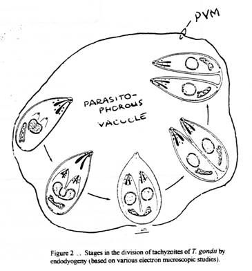
invade the cells of the intestinal epithelium and divide by a process called endodyogeny.
Endodyogeny is the development of 2
daughter parasites within a mother cell by a process of intracellular binary
fission.
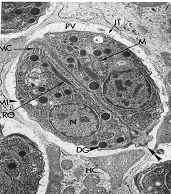
Tachyzoites in endodyogeny
by TEM
MC, mother cell; PV,
parasitophorous vacuole; HC, host cell; RO, rhoptry; M, mitochondrion; MC,
micronemes; DG, dense granule; IT, intravacuolar tubules; MI, micropore
The inner membrane of the mother cell disappears and the
inner membrane of the daughter cell joins the other membrane of the mother
cell. If held together at the posterior end with repeated cycles of endodyogeny
a rosette of crescent-shaped parasites is formed. After several cycles
of asexual reproduction gamogony begins: the intracellular stages do not undergo endodyogeny, but instead
differentiate into gametocytes, and fertilization occurs.Two walls are laid
down around the zygote to form the oocyst, and
unsporulated oocysts are found in the feces in less than a week. Sexual
stages are produced only in the definitive feline host!

Unsporulated oocysts in cat feces. F. Isospora
felis and T. Toxoplasma gondii
Sex is not determined genetically in that a single tachyzoite or sporozoite can go on to form either male or female gametocytes. A
single tachyzoite or sporozoite infecting a cat gives rise to normal oocysts in
the cat. Therefore, a physiologic trigger for gametocytogenesis must exist and
it must involve the cat intestinal epithelium since gametocytes never form in
vitro. The cue for the microgamete finding the macrogamete is unknown.)
Simultaneously, the
bradyzoites penetrate the lamina propia of the cat intestine where they
multiply as crescent-shaped tachyzoites.
Aggregates of tachyzoites are enclosed
within a pseudocyst of parasite and host origin. Tachyzoites can enter almost
any nucleated cell, and multiply until the host cell is filled with the next
generation of tachyzoites. In the process the host cell is killed. This cycle
of parasite replication and host cell
death may result in microscopic centers of
necrosis.
Within hours after infection the tachyzoites are engulfed by phagocytic cells and
move via the lymph and blood to
extra-intestinal sites where
endodyogeny continues. The host usually
overcomes this acute phase of infection, and then the chronic phase
begins: the parasites (now called bradyzoites), within a cyst, divide slowly by endodyogeny, and persist in
the tissues for months if not for the life of the cat. Thus, if a cat is fed
bradyzoites development of Toxoplasma occurs in both the intestine
and the tissues. The tissue cysts usually cause no host reaction. If a cat is fed bradyzoites in meat there will be oocysts in the feces in
<10 days, whereas if fed tissue containing tachyzoites oocysts are shed
within 2 weeks, and if fed oocysts then oocysts are found in the feces in
>18days.
Fewer than 50% of
cats shed oocysts if fed tachyzoites or oocysts, but 100% shed oocysts if fed
bradyzoites. Clearly, Toxoplasma is a
coccidian parasite that has evolved mechanisms for infecting its definitive
host, the cat, and it utilizes its prey
(rodents and birds), as well as oocysts, to accomplish its dispersal
from one host to another.
In humans, after
ingestion of oocysts, tachyzoites are found during the first 2 weeks, and constitute the acute phase of the
infection.
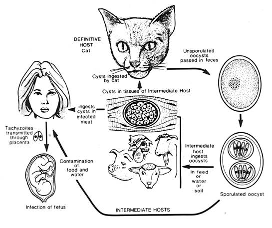
The life
cycle of T. gondii
*Formation of Cysts with Brayzoites
In the mouse
development may be more rapid than in humans but
it is clear that tachyzoites are disseminated
throughout the body tissues via the blood and
lymph and this takes 4 days. Five days after
oocyst ingestion there are tissue cysts in the
intestine, and by 8 days tissue cysts are found in
the brain. The tissue cyst is pepsin-resistant and
develops from the parasitophorous vacuolar
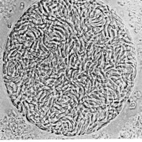
membrane. A cyst may contain up to 3000 bradyzoites, and most cysts occur in the muscle and brain and
can persist for years. This is the chronic phase
of infection. If humans or other carnivores (not
cats!) eat meat containing cysts with bradyzoites
then these stages emerge and invade cells (other
than intestinal cells) and they divide as
tachyzoites first leading to an acute disease,
then to a chronic disease.
No oocysts are formed!
 T
T
Cyst
with bradyzoites