Life Cycle and Transmission
The life cycle of G. duodenalis is relatively simple and alternates between the actively metabolizing motile stage, the trophozoite, and the dormant, environmentally resistant cyst.
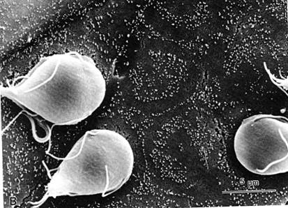
The trophozoite resembles a pear cut in half longitudinally, is binucleate, ranges in size from 10 to 15mm in length and 6-10mm in width and 2-4mm thick, and swims in the intestinal fluid and tethers itself to the mucus strands by means of 4 pairs of flagella.

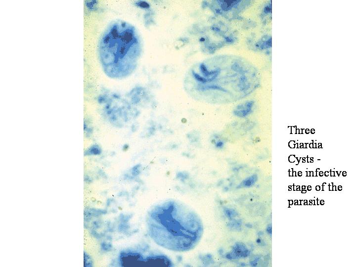
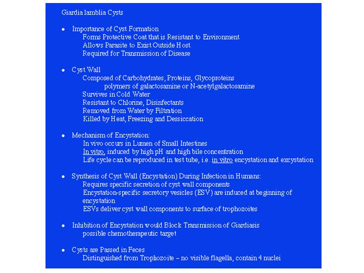
The cyst form is 5-8mm and is surrounded by a wall that is 0.3mm thick. The outer wall consists of many filaments, whereas the inner wall contains outer and inner cyst membranes separated by a thin layer of cytoplasm. The major sugar of the outer cyst wall is N-acetylgalactosamine (GalNAc) plus galactose, and the cyst appears not to contain chitin. In addition, there are two cyst wall proteins that have been characterized and whose genes have been sequenced.
The trophozoites live
in the small intestine (mid-jejunum) attached to the surface of the enterocytes
by the ventral adhesive disc, dividing every 9-12hr. In 10 days there could be
1 million parasites and in two weeks a billion Giardia. Trophozoites may
detach intermittently, but
this is of minimal duration since it places the parasite in the position of
being swept downstream by peristalsis.
In the
mid-jejunum they are exposed to a
cholesterol-rich environment (from both food and bile). Cholesterol is absorbed
primarily in the jejunum whereas bile acid absorption occurs in the ileum.
Bacterial populations also decrease the amount of bile and chemically alter the
cholesterol thereby decreasing its availability for the trophozoites. Thus, as
trophozoites pass to the posterior part of the small intestine they are exposed
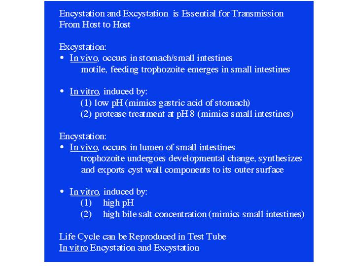
to increasedlevels of bile and higher pH, and this triggers
encystation. Within 5 hr encystation-specific
antigens can be found in the endoplasmic reticulum,
and are packaged into
encystation-specific vacuoles, which appear in 6-18 hr, and these are
transported to the forming cyst wall. Cyst formation begins on the dorsal
surface, there is a change in shape--flattened pear-shaped to spherical--the
flagella
curl up inside cyst wall and the trophozoite becomes
enclosed in the wall. Nuclei divide during encystment, the disc disorganizes, and a quadrinucleate cyst is discharged with the
feces. It is not clear whether the cyst is infective immediately after passage,
however, there is some evidence to show
that it may take 7 days before full infectivity can occur.
Summary of Giardia Life Cycle:
• Infection is acquired by ingestion of mature cysts.
•Cysts have rigid cell walls and are non-dividing. They can survive in water
and are resistant to dessication.
• Cysts pass through the stomach, where they are exposed to gastric acid/low pH
and emerge in the lower stomach or upper small intestine as a trophozoite.
• The trophozoite colonize the small intestine and are the disease-causing stage.
• The trophozoite adheres to the epithelia, using a ventral adhesive disc, and can
cause malabsorption from the intestines and diarrhea.
• Trophozoites that migrate to the distal small intestine undergo a process
of encystation, triggered by high pH and bile salts. Encystation requires
the synthesis of a cell wall.
• Cysts are passed in the feces of the host and mature and survive externally
