
Infection
is via the fecal-oral route and C. parvum can infect epithelial cells of
the gastrointestinal and respiratory tracts, and the biliary and pancreatic
ducts. Cryptosporidium is so named because the oocyst contains 4 naked
sporozoites i.e. the sporocyst is either concealed or absent. (FIGURE 1).
http://www.ksu.edu/parasitology/basicbio
http://www.biosci.ohio-state.edu/~parasite/cryptosporidium.html
Figure 1.
Diagrammatic representation of C. parvum life cycle (after Fayer)
Like that of other coccidia (see Isospora
belli) the oocyst wall of C. parvum has distinct inner and outer
layers with a suture at one end. (FIGURE 2A).
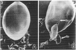
Figure
2. A. Intact oocyst; B. Sporozoites emerging from oocyst
Su, suture; Sp, sporozoite
The
infection is acquired by the ingestion of water or food contaminated with
mature oocysts passed in the feces. Once in the intestine, the
trypsin-sensitive suture dissolves during excystation, thereby opening the wall
through which the sporozoites leave the oocyst. (FIGURE 2B) The sporozoites
attach to the enterocytes, and by means of the apical organelles—rhoptries and
micronemes--entry into the enterocyte is gained (FIGURE 3).

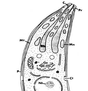
Figure 3.
A. Sporozoite by TEM B. Sporozoite
(diagrammatic)
Ec,
electron dense collar; Pl, pellicle; Mn, micronemes; Rb, residual body; Rh,
rhoptry; Art 1,2, apical rings; Im, inner membrane; Mt, microtubule;Gc, Golgi
complex; C, conoid; Ct, cytostome; Pr, polar rings; NI, nucleolus
In addition to these apical organelles the
sporozoites also have a pellicle, electron dense granules, ribosomes,
subpellicular microtubules, nucleus and apical rings, but lack mitochondria,
polar rings, micropore and conoid. Within the enterocyte the trophozoite (and
schizont) are located extracytoplasmically, just under the brush border
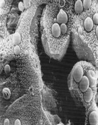
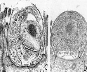
Figure 4.
Scanning EM of C. parvum Transmission EM of
C. parvum
Ec,
electron dense collar; FI, fibrous layer; El, electron dense layer (From Fayer
at al)
Two schizogonic cycles take place: type I schizonts produce 8 merozoites, and type II produce 4 merozoites. The latter invade fresh enterocytes and undergo gametogony to produce macro and microgametes. There is fusion of gametes, and the formation of one of two kinds of oocysts: thin-walled and thick-walled. Sporogony takes place within the oocyst and gives rise to 4 sporozoites. The thick-walled oocysts are passed out with the feces, whereas the thin-walled oocysts, which are already infective, can excyst within the lumen of the intestine, resulting in autoinfection. (FIGURE 5).
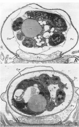
Figure 5. TEM of oocysts. A. Thin-walled and B. Thick walled
Ow, outer
wall; Lb, lipid body; Sp, sporozoite; Ec, electron dense collar; Fo, feeder, organelle;
Pv, parasitophorous vacuole; Su, suture
In this way
an infection can begin anew without ingestion of oocysts. The time after ingestion of infective oocysts to
complete the life cycle and to have new oocysts excreted is 4-22 days in humans
and 2-7 days for calves.