
Morphology and Life Cycle
Hemoflagellates have the typical cell components of a eukaryote: nucleus, microtubules (cytoskeleton and flagellar), endoplasmic reticulum, Golgi apparatus and a single mitochondrion. In addition, they have another body, the kinetoplast, at the base of the flagellum. It was originally believed that this body was some kind of cellular motor that caused the flagellum to move, hence the terminology “kineto” in the order name. Now we know that the kinetoplast is an extension of the mitochondrion and contains the mitochondrial genome. Hemoflagellates have several morphologic forms: amastigote, epimastigote, trypomastigote and promastigote.
1) The TRYPOMASTIGOTE is found in the bloodstream of infected vertebrates. It is a non-dividing form that is infectious for the reduviid bug. It often has a characteristic ‘C’ shape in Giemsa stains of bloodsmears. The greek suffix "mastigote" means ‘whip-like’ referring to the single flagellum, which emerges through a flagellar pocket and runs the whole length of the cell. The large kinetoplast is found at the posterior end of the cell. The METACYCLIC TRYPOMASTIGOTE is found in bug feces and is the infectious stage for vertebrates.
 Blood (right) and Metacyclic (left) trypomastigotes |
|
2) The AMASTIGOTE is the intracellular dividing form in the cytoplasm of vertebrate cells. It is a round/oval-shaped cell with no protruding flagellum.

Taken from Mackie, Hunter and Worth, Manual of Tropical Medicine, W.B. Saunders Co., Phila. 1945
3) The EPIMASTIGOTE is found in the intestinal tract of the insect vector. In this form, the kinetoplast is found anterior and adjacent to the nucleus (in greek, "epi" means ‘upon’). The flagellum emerges in the middle of the cell. Below are an epimastiogote (left) and a metacyclic trypomastigote (right) in bug feces.

Life Cycle
T. cruzi is a pathogen of humans and other vertebrates. It can be found in the bloodstream and inside macrophages and muscle cells. The most common means of transmission between vertebrate hosts is through several species of triatomine bugs (order Hemiptera and family Reduviidae). These triatomine or reduviid bugs are commonly called “cone-nose bugs,” “assassin bugs”, “kissing bugs” or “barbiero (barber) bugs” because they tend to bite on the face. The bite is painless, but the saliva produces an allergic response which causes the person to scratch.
The principal reservoirs for human infections are domestic dogs and cats. Opossums and armadillos, which are infected for life and infect triatomines, which can invade houses or infect domestic animals, play a major role in joining the sylvatic and domestic cycles
The natural (vector-borne)
cycle of transmission
When a triatomine bug bites an infected individual it ingests trypomastigotes—it can take as few as 10 parasites to infect 63% of bugs and even five can infect a bug. Within the gut lumen, trypomastigotes differentiate into epimastigotes, these migrate to the midgut where they multiply. The epimastigotes, which are resistant to midgut proteases, migrate to the hindgut, attach to rectal glands and differentiate into metacyclic trypomastigotes. The trigger for differentiation is unknown, but in vitro a solution mimicking insect urine + proline promotes attachment to the substratum and differentiation. From ingestion of the blood-borne trypomastigotes to the development of metacyclic trypomastigotes in the gut takes 7 days.
The form of T. cruzi that is ingested and infectious for the reduviid is the bloodstream trypomastigote. In the intestines of the bug, the trypomastigote differentiates into the epimastigote form, which attaches to the intestinal wall and divides by binary fission. In the hindgut (posterior station), the epimastigotes differentiate into metacyclic trypomastigotes which are passed out in the feces. The common reduviid vectors such as R. prolixus and Panstrongylus megistus defecate during the bloodmeal, allowing efficient infection by the parasite. Contaminated feces is pushed by scratching into either the puncture wound left after feeding or into mucous membranes, commonly those of the eye. The metacyclic trypomastigote enters cells at the site of inoculum and differentiate into the amastigote form. Amastigotes divide until the cell bursts, whereupon they are released and infect neighboring cells. Within some infected cells, the amastigotes differentiate into trypomastigotes that eventually get into the bloodstream from where they have access to infect muscle cells. Inside muscle cells, they replicate as amastigotes, which can differentiate into trypomastigotes and gain access back into the bloodstream, where they must be ingested by a reduviid bug to continue the life cycle.
There are several genera of Reduviidae that can transmit the protozoan. Triatoma infestans, as its name suggests, frequently invades homes (mud and stick huts) and is responsible for what is termed the domestic transmission cycle. Other reduviids, such as Rhodnius prolixus, reside in rural settings or forests, they are associated with the sylvatic cycle of transmission, which includes many wild animals as vertebrate hosts. The natural hosts include dogs, cats, guinea pigs, opossums, foxes, squirrels, armadillos, porcupines, rats, mice and bats. T. cruzi is infectious to a wide range of vertebrate hosts and is considered to be a zoonosis that only recently has spread to humans.
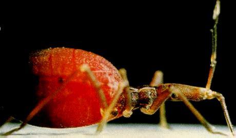
One happy triatomid !
Contaminated feces (there can be 3500 trypomastigotes per microliter of excreta) are pushed by scratching into either the puncture wound left after feeding or into mucous membranes, commonly those of the eye. The metacyclic trypomastigotes that enter the wound by contamination are phagocytized by macrophages, escape from phagolysosome come to lie in the cytoplasm, and differentiate into amastigotes. Amastigotes multiply by binary fission until the cell is filled, bursts, and the released amastigotes can then infect neighboring cells. Within some infected cells the amastigotes differentiate into trypomastigotes that eventually leave the cell, and enter the bloodstream from where they have access to infect muscle cells. Inside the muscle cells, they replicate as amastigotes, which can in turn, differentiate into trypomastigotes and gain access back into the bloodstream where they must be ingested by a reduviid bug to continue the cycle.
If trypomastigotes in the blood are inoculated into another vertebrate they persist in the bloodstream for several days and then invade cells. If however metacyclic trypomastigotes (in bug feces) are inoculated they enter host cells at the site of inoculation, multiply at this site, and then they move into the bloodstream, and subsequently to other tissues.
The life cycle can be obtained in vitro for experimental purposes; the easiest form to culture is the epimastigote, which grows in LIT (Liver Infusion Tryptose) medium at 28oC. These can be induced to differentiate into trypomastigotes in TAU (Triatomine Artificial Urine) medium. Trypomastigotes can infect tissue culture cells (and experimental animals such as mice) where they replicate as amastigotes, and even differentiate into trypomastigotes.
Questions:
1. How are people infected with T. cruzi?
2. What behavior of the insect host is correlated with infectivity?
3. What morphological feature of the cell is used to distinguish different life cycle forms? What are the three major forms in the life cycle of T. cruzi?